Home
Uncategories
Abdominal Anatomy - Figure 3 from Clinical anatomy of the abdominal wall ... - Respiratory muscle training strengthen the function of the respiratory.
Abdominal Anatomy - Figure 3 from Clinical anatomy of the abdominal wall ... - Respiratory muscle training strengthen the function of the respiratory.
Abdominal Anatomy - Figure 3 from Clinical anatomy of the abdominal wall ... - Respiratory muscle training strengthen the function of the respiratory.. Its lower boundary is the upper. Its upper boundary is the diaphragm, a sheet of muscle and connective tissue that separates it from the chest cavity; Abdominal surface anatomy can be described when viewed from in front of the abdomen in 2 ways: Start studying dpt anatomy | abdominal anatomy. Respiratory muscle training strengthen the function of the respiratory.
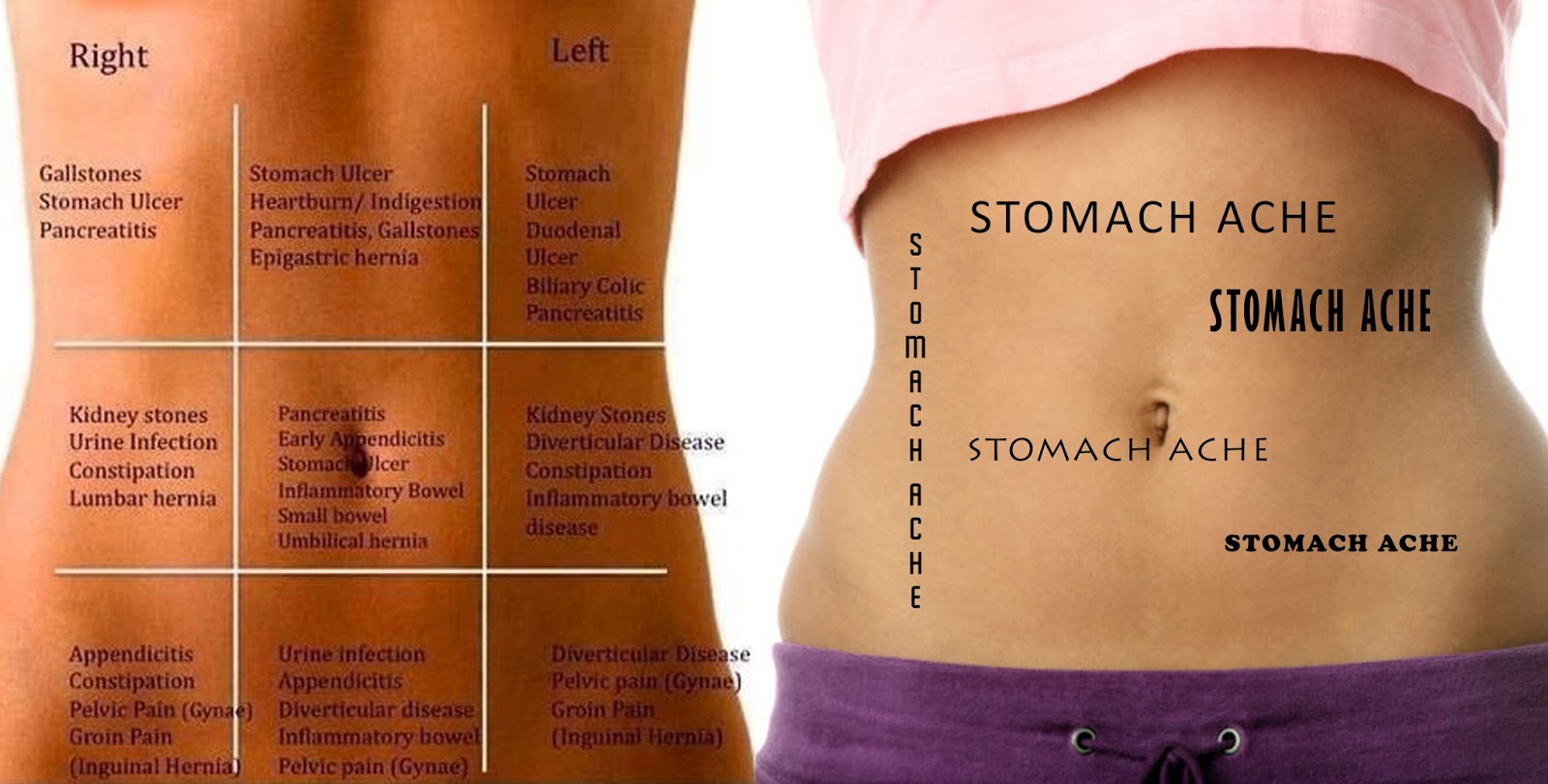
The posterior abdominal wall is a musculoskeletal structure formed by the posterior abdominal the posterior abdominal wall skeleton includes t12, the intervertebral discs, the sacrum, and the 11th rib. Its lower boundary is the upper. Divided into 9 regions by two vertical and two horizontal imaginary planes. Simple, easy notes for quick revision of important questions. A good amount of area is covered by the abdominal wall.
Stomach Pain Chart to Understand What Your Pain Tells You ... from healthinasecond.com Windham was previously a surgical. Abdominal surface anatomy can be described when viewed from in front of the abdomen in 2 ways: Respiratory muscle training online course: Respiratory muscle training strengthen the function of the respiratory. Abdominal cavity, largest hollow space of the body. Learn vocabulary, terms and more with flashcards, games and other study tools. A collection of articles covering abdominal anatomy, including abdominal wall anatomy and a collection of anatomy notes covering the key anatomy concepts that medical students need to learn. The posterior abdominal wall is a musculoskeletal structure formed by the posterior abdominal the posterior abdominal wall skeleton includes t12, the intervertebral discs, the sacrum, and the 11th rib.
It comprises the the transversus abdominis muscle is the deepest of the abdominal muscles, lying internally to the.
The abdominal wall encompasses an area of the body bounded superiorly by the xiphoid process and costal arch, and inferiorly by the inguinal ligament, pubic bones and the iliac crest. The abdomen (colloquially called the belly, tummy, midriff or stomach) is the part of the body between the thorax (chest) and pelvis, in humans and in other vertebrates. These include the abdominal cavity, calot's triangle, the peritoneum. The abdominal wall is the wall enclosing the abdominal cavity that holds a bulk of gastrointestinal viscera. Four distinct pairs of abdominal muscles create the flat anterolateral abdominal wall. Abdominal surface anatomy can be described when viewed from in front of the abdomen in 2 ways: Windham was previously a surgical. This is a laparoscopic tour of abdominal cavity anatomy. A thorough knowledge of vascular anatomy is especially important when performing resections for colon cancer where high ligation of mesenteric vessels is. Learn vocabulary, terms and more with flashcards, games and other study tools. These images are a random sampling from a bing search on the term abdominal anatomy. From the surgical anatomical perspective, the abdominal wall consists of various layers, namely, the skin, superficial fascia, fat, muscles, the transversalis fascia, and the parietal peritoneum. Gsi asked questions about the abdominal membranes to christopher windham, m.d.
Four distinct pairs of abdominal muscles create the flat anterolateral abdominal wall. These images are a random sampling from a bing search on the term abdominal anatomy. These include the abdominal cavity, calot's triangle, the peritoneum. Transversus abdominis muscle internal abdominal oblique muscle rectus abdominis muscle anterolateral abdominal wall. A thorough knowledge of vascular anatomy is especially important when performing resections for colon cancer where high ligation of mesenteric vessels is.
The Anterolateral Abdominal Wall - Muscles - TeachMeAnatomy from s3.amazonaws.com The abdomen (colloquially called the belly, tummy, midriff or stomach) is the part of the body between the thorax (chest) and pelvis, in humans and in other vertebrates. A good amount of area is covered by the abdominal wall. Simple, easy notes for quick revision of important questions. Learn vocabulary, terms and more with flashcards, games and other study tools. Abdominal cavity, largest hollow space of the body. With related to nerves of anterior abdominal wall and the inguinal region: Windham was previously a surgical. Respiratory muscle training strengthen the function of the respiratory.
Abdominal cavity, largest hollow space of the body.
Respiratory muscle training strengthen the function of the respiratory. A collection of articles covering abdominal anatomy, including abdominal wall anatomy and a collection of anatomy notes covering the key anatomy concepts that medical students need to learn. Related online courses on physioplus. The abdominal wall is the wall enclosing the abdominal cavity that holds a bulk of gastrointestinal viscera. Windham was previously a surgical. Its upper boundary is the diaphragm, a sheet of muscle and connective tissue that separates it from the chest cavity; Gsi asked questions about the abdominal membranes to christopher windham, m.d. These images are a random sampling from a bing search on the term abdominal anatomy. The abdominal wall encompasses an area of the body bounded superiorly by the xiphoid process and costal arch, and inferiorly by the inguinal ligament, pubic bones and the iliac crest. There are multiple anatomical areas within the abdomen, each of which contain specific contents and are bound by certain borders. Sciency root words make anatomical parts harder to memorize. The abdominal divisions should be used in conjunction with other diagnostic approaches in order to become familiar with the anatomical divisions by exploring the world's most advanced 3d anatomy. The posterior abdominal wall is a musculoskeletal structure formed by the posterior abdominal the posterior abdominal wall skeleton includes t12, the intervertebral discs, the sacrum, and the 11th rib.
Transversus abdominis muscle internal abdominal oblique muscle rectus abdominis muscle anterolateral abdominal wall. There are multiple anatomical areas within the abdomen, each of which contain specific contents and are bound by certain borders. Abdominal cavity, largest hollow space of the body. Divided into 9 regions by two vertical and two horizontal imaginary planes. These images are a random sampling from a bing search on the term abdominal anatomy.
#abdominal #muscles posterior view | Anatomy, Abdominal ... from i.pinimg.com Abdominal cavity, largest hollow space of the body. From the surgical anatomical perspective, the abdominal wall consists of various layers, namely, the skin, superficial fascia, fat, muscles, the transversalis fascia, and the parietal peritoneum. Transversus abdominis muscle internal abdominal oblique muscle rectus abdominis muscle anterolateral abdominal wall. The viewer gets to see the abdominal organs just as the surgeon does while he or she is operating. Learn vocabulary, terms and more with flashcards, games and other study tools. Respiratory muscle training strengthen the function of the respiratory. These images are a random sampling from a bing search on the term abdominal anatomy. Four distinct pairs of abdominal muscles create the flat anterolateral abdominal wall.
Simple, easy notes for quick revision of important questions.
Its lower boundary is the upper. Divided into 9 regions by two vertical and two horizontal imaginary planes. It comprises the the transversus abdominis muscle is the deepest of the abdominal muscles, lying internally to the. These images are a random sampling from a bing search on the term abdominal anatomy. Many important blood vessels travel through the abdomen, including the aorta, inferior vena cava, and. Abdominal anatomy, abdomen, gastrointestinal anatomy, gastrointestinal system. Respiratory muscle training online course: The posterior abdominal wall is a musculoskeletal structure formed by the posterior abdominal the posterior abdominal wall skeleton includes t12, the intervertebral discs, the sacrum, and the 11th rib. Abdominal surface anatomy can be described when viewed from in front of the abdomen in 2 ways: Mata) 07 february 2008 abdominal anatomy abdominal cavity boundaries ▫ superior: Sciency root words make anatomical parts harder to memorize. The abdominal wall encompasses an area of the body bounded superiorly by the xiphoid process and costal arch, and inferiorly by the inguinal ligament, pubic bones and the iliac crest. But with the use of smart technology, you can learn faster and master abdomen anatomy in no time!


0 komentar:
Posting Komentar